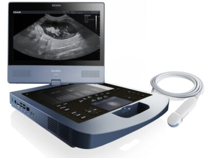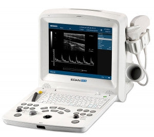
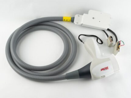
GE VOLUSON E8 Refurbished WITH 3 Probes
Refurbished with 3 Probes included
90 Day warranty
GE Voluson E8 Ultrasound Review
The GE Voluson E8 has high-quality images in all settings, but the system is most impressive when you are viewing its HDlive high-resolution 4D images. HDlive is GE’s advanced 4D imaging in high definition. The resolution and detail of HDlive versus traditional 4D images are breathtaking and can improve the ultrasound’s efficiency in clinical applications. The difference can be compared to switching from an analog TV to an HD TV. Since GE launched HDlive in 2012, several other manufacturers have created their own versions of HD 4D imagery. However, the HDlive featured on the Voluson E8 is still the most popular. The detail, shading options, and fast frame rates of the Voluson E8 keep this ultrasound at the top of the pack for 4D ultrasounds. The enhanced 4D resolution on the Voluson E8 also enables obstetricians to have a better volume assessment of the first-trimester anatomy as well as vascular structures. The 4D images and movies generated by the Voluson E8 can also be exported in a variety of formats, making this one of the highest quality and most effective ultrasound systems for women’s health applications.
GE Voluson E8 vs. Other GE Ultrasound Machines
The GE Voluson E8 ultrasound is the successor to the Voluson 730 Expert, previously the most popular and well-known 4D ultrasound machine. The Voluson E8 improves upon the Voluson 730 in many aspects, including screen size, resolution, speed, weight, and features. Both the GE Voluson E6 and the Voluson S8 are slightly less powerful and less expensive than the Voluson E8. The Voluson S8 is a more recent design and therefore smaller and lighter than the larger Voluson E8. The Voluson S8 has half the channels and a smaller selection of transducers than the Voluson E8, however. The Voluson E10 ultrasound is a newer and more powerful system than the Voluson E8 with a larger monitor, enhanced features, and more available transducers, but all of that comes at a higher price from $47,000 up to $51,000 or more. The GE Logiq E9 is similar in price to the Voluson E8 and can perform all applications well, whereas the Voluson E8 is focused primarily on women’s health and 4D applications. If you will be performing general imaging or vascular or cardiac scans on adults, then the Logiq E9 is a better choice. Otherwise, the Voluson E8 is worth your consideration.
GE Voluson E6 vs. Voluson E8
The GE Voluson E6 looks identical to the Voluson E8 from the exterior, except for the nameplate. The Voluson E6 is essentially a Voluson E8 with fewer features and probes available as well as only half the channels. Because of its lower number of channels, the GE Voluson E6 is actually closer in performance to the newer GE Voluson S8 than the Voluson E8. For this reason, the Voluson E8 is a better seller than the Voluson E6. In the opinion of many physicians, the Voluson E8 is worth the extra cost over the Voluson E6 because of its superior performance.
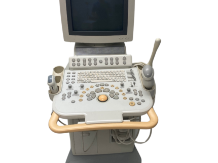
GE Voluson E8 Standard Features
19” LCD monitor on fully articulating arm
10.4” LCD touch panel
Automatic Tissue Optimization
Coded Harmonic Imaging with Pulse Inversion Technology
Coded Excitation (CE)
FFC – Focus Frequency Composite
HD-Flow
B-Flow
Virtual Convex and Wide Sector
Tissue Doppler
XTD (Extended View or Panoramic Imaging Mode)
SRI II (Speckle Reduction Imaging)
CrossXBeamCRI* (Compound Resolution Imaging)
TUI (Tomographic Ultrasound Imaging, or Multi-Slice Imaging)
SonoNT
SonoIT
SonoBiometry
SonoRender Start
Scan Assistant
DICOM™ (Verify, Print, Store, Modality Worklist, Structured Reporting, MPPS, Media Exchange)
Static 3D Mode:
B Mode only
B + Power Doppler Mod
B + CFM Doppler Mod
B + HD-Flow Mod
B + CRI
Focus and Frequency Composite (FFC)
High-Resolution Zoom
Pan Zoom
Steering
Virtual Convex
Wide Angle on Endovaginal Probes
BetaView
Patient Information Database
Image Archive on a Hard Drive (via Raw-Data File, DICOM File both Single and Multiframe)
Image Archive to CD-R(W), DVD +/-
Export Images as BMP, TIFF, JPEG, AVI, MOV, DCM. MPEG4
3D/4D Data Compression (Lossy/Lossless)
Inversion
Real-Time Automatic Doppler Calculations
Hard Drive 500 GB (BT13), 450GB for Data Storage
OB Measurements, Calculations, and Reports
GYN Measurements, Calculations, and Reports
Vascular Measurements, Calculations, and Reports
Cardio Measurements, Calculations, and Reports
Abdominal Measurements, Calculations, and Reports
Multigestational Calculations
GE Voluson E8 Ultrasound Technology Definitions
Coded Excitation (CE): This feature on the Voluson E8 improves image resolution and penetration in the far-field. This allows the use of a higher frequency on technically difficult patients.
Wide Sector: Standard on the Voluson E8, this widens the sector probe’s imaging field of view.
SRI II: A nonlinear diffusion filtering technique that improves image quality in real-time by reducing speckles.
CrossXBeam CRI: This core technology on the GE Voluson E8 uses compound resolution imaging to improve border and image clarity.
TUI: This is a new visualization mode for 3D and 4D data sets on the Voluson E8. The data is presented as slices through the data set that are parallel to each other. An overview image, which is orthogonal to the parallel slices, shows which parts of the volume are displayed in the parallel planes. This method of visualization is consistent with the way other medical systems such as CT or MRI, present the data to the user. The distance between the different planes can be adjusted to the requirements of the given data set. In addition, it is possible to set the number of planes. The planes and the overview image can also be printed to a DICOM printer, for easier comparison
of the ultrasound data with CT and/or MRI data.
SonoNT: This Voluson E8 feature allows for semi-automatic Nuchal Translucency measurements.
SonoIT: Sonography based Intracranial Translucency is a system supported measurement.
Starting from the routinely used midsagittal view of the fetal face, obtained for assessment of the Nuchal Translucency and nasal bone, the ultrasound system uses a semiautomated mode to measure the anterior-posterior diameter of the fourth ventricle recognizable as intracranial translucency. The workflow is identical to SonoNT.
SonoBiometry: The Voluson E8 provides an alternative to the common fetal biometry measurements. It provides system suggested measurements for BPD, HC, AC, and FL which need to be confirmed by the user or can be changed manually.
SonoRender Start: This feature on the Voluson E8 speeds up the acquisition of the fetal face in 4D.
Scan Assistant: Automated step-by-step help for those new to scanning.
Focus and Frequency Composite (FFC): A Voluson E8 technology that utilizes two different transmission frequencies and two different focal ranges in the 2D image. This function combines a
low frequency to increase the penetration and higher frequency to keep a high resolution. It reduces speckle and artifacts in the 2D image to facilitate the examination of difficult-to-scan patients.
Beta View: Allows for the adjustment of the Volume O-Axis position of 3D probes in 2D mode. The green line in the displayed symbol indicates the position of the acoustic block. The “+” and “-“ define the corresponding sweep direction on the touchscreen.